Visual cortex
Brain region processing visual information from the eyes.
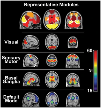
The visual cortex is the area of the cerebral cortex in mammals that processes visual information. Located in the occipital lobe at the rear of the head, it receives sensory input from the eyes via the lateral geniculate nucleus in the thalamus. The primary visual cortex (V1), also known as the striate cortex or Brodmann area 17, is the first cortical area to receive this input, and extrastriate areas (V2, V3, V4, V5) handle further processing. Both hemispheres contain a visual cortex, with the left hemisphere receiving signals from the right visual field and the right hemisphere from the left visual field.
- location
- Occipital lobe, posterior pole of the brain
- key_components
- Primary visual cortex (V1) and extrastriate areas (V2, V3, V4, V5)
- neuron_count
- Approximately 140 million neurons per hemisphere in adult humans
- average_volume
- About 5400 mm³ per hemisphere in adult humans
- blood_supply
- Calcarine branch of the posterior cerebral artery
- functional_pathways
- Ventral stream (form recognition) and dorsal stream (motion and spatial location)
Lore & Background
The visual cortex is organized retinotopically, meaning neighboring cells in V1 have receptive fields corresponding to adjacent portions of the visual field. Neurons in V1 fire action potentials when visual stimuli appear within their receptive field, and they exhibit tuning—for example, a V1 neuron may respond best to a vertical stimulus. In higher visual areas like the inferior temporal cortex, neurons have more complex tuning, such as responding only to a specific face. V1 is characterized by a laminar organization with six layers; layer 4 receives most input from the lateral geniculate nucleus and is further divided into sublaminae that process magnocellular and parvocellular pathways.
Reader's Guide
The visual cortex is fundamental to understanding how the brain processes visual information. V1 is the most studied visual area, highly specialized for pattern recognition and processing static and moving objects. It transmits information via two primary pathways: the ventral stream (associated with form recognition and object representation) and the dorsal stream (associated with motion, object location, and guiding actions). The what vs. where account of these pathways was first described by Ungerleider and Mishkin, with later extensions by Goodale and Milner suggesting the ventral stream is critical for perception and the dorsal stream for visual control of actions. Studies on visual illusions, such as the Ebbinghaus illusion, have produced conflicting evidence on whether action and perception systems are equally affected. V1 also exhibits plasticity, allowing functional and structural changes from sensory experience. The size of V1, V2, and V3 can vary three-fold, a difference partially inherited.
Did You Know?
- The primary visual cortex is also known as the striate cortex due to the line of Gennari, a visible stripe of myelinated axons.
- Each hemisphere's V1 receives information from the contralateral visual hemifield via the ipsilateral lateral geniculate nucleus.
- The average number of neurons in the adult human primary visual cortex per hemisphere is about 140 million.
- The volume of V1 in adult humans varies widely, from 3185 to 7568 mm³ in the left hemisphere.
More in Sensory Systems 25-35
Elsewhere in the Sensory Systems universe
Spotted an error? Know more?
This is a living reference — every entry is fact-audited, and reader corrections feed straight into our audit queue. Suggest an edit · See this site's audit record